File:Polyomavirus.jpg

Original file (2,419 × 1,738 pixels, file size: 429 KB, MIME type: image/jpeg)
Summary[edit]
| Summary | |
|---|---|
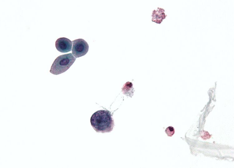
| Description | Micrograph showing a polyomavirus infected cell. Urine cytology specimen.
Features of polyomavirus infected cells (also known as Decoy cells): Usually 2X the size of a basal urothelial cell nucleus. Single cells - important feature. Scant "degenerative-appearing" cytoplasm. High NC ratio. Intranuclear inclusions - key feature. Central smudging (or "wash-out") of the chromatin/"Ground glass" chromatin. Surrounded by clear halo just deep to the nuclear membrane. Nuclear membrane clumping. See also Image:Polyomavirus 2.jpg |
| Source | Wikimedia Commons file page |
| Author | Nephron |
| Permission | See original Commons license details. |
Licensing[edit]
Creative Commons Attribution-ShareAlike 3.0 Unported (CC BY-SA 3.0)
This file is licensed under the Creative Commons Attribution-ShareAlike 3.0 license.
Official license: CC BY-SA 3.0
License page: CC BY-SA 3.0
Original attribution and file history: Wikimedia Commons
File history
Click on a date/time to view the file as it appeared at that time.
| Date/Time | Thumbnail | Dimensions | User | Comment | |
|---|---|---|---|---|---|
| current | 03:35, 5 June 2026 | | 2,419 × 1,738 (429 KB) | Maintenance script (talk | contribs) | == Summary == Importing file |
You cannot overwrite this file.
File usage
The following 2 pages use this file:
