File:Ramos cell trogocytosis.png
From WikiMD's WELLNESSPEDIA

Size of this preview: 800 × 487 pixels. Other resolution: 1,183 × 720 pixels.
Original file (1,183 × 720 pixels, file size: 287 KB, MIME type: image/png)
Summary[edit]
| Summary | |
|---|---|
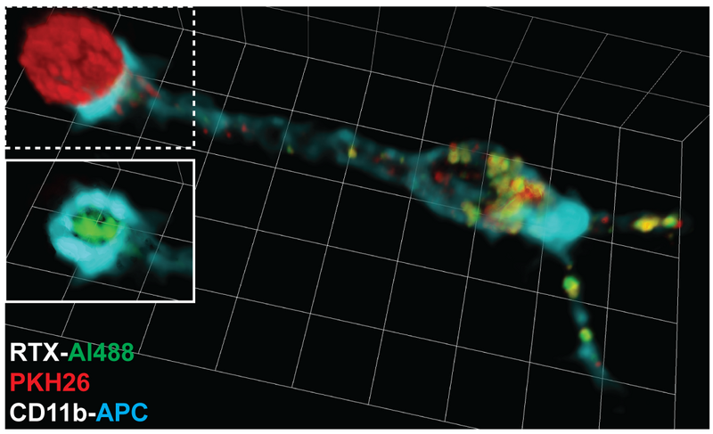
| Description | Volumetric reconstruction from confocal slices of a Ramos-RAW cell interface. RTX-Al488-coated (green), PKH26-labelled Ramos cells (red) were incubated with RAW cells for 45 minutes at 37°C. RAW cells were labelled with anti-CD11b-APC (cyan). The RAW cell has extensively trogocytosed both RTX and PKH26. Inset shows the dotted area above it without the PKH26 channel overlaid, revealing the concentration of RTX-Al488 at the cell-cell interface, otherwise depleted from the rest of the Ramos cell. Trogocytosis reaction was halted by fixation 45 min after co-incubation. Ramos cells are approximately 12 µm in diameter. |
| Source | Wikimedia Commons file page |
| Author | Theodore Pham, Patricia Mero, James W. Booth. |
| Permission | See original Commons license details. |
Licensing[edit]
License: CC BY 2.5
License page: CC BY 2.5
Original attribution and file history: Wikimedia Commons
File history
Click on a date/time to view the file as it appeared at that time.
| Date/Time | Thumbnail | Dimensions | User | Comment | |
|---|---|---|---|---|---|
| current | 22:30, 8 June 2026 | | 1,183 × 720 (287 KB) | Maintenance script (talk | contribs) | == Summary == Importing file |
You cannot overwrite this file.
File usage
The following page uses this file:
