File:Renal oncocytoma2.jpg
Original file (1,952 × 1,268 pixels, file size: 758 KB, MIME type: image/jpeg)
Summary[edit]
| Summary | |
|---|---|
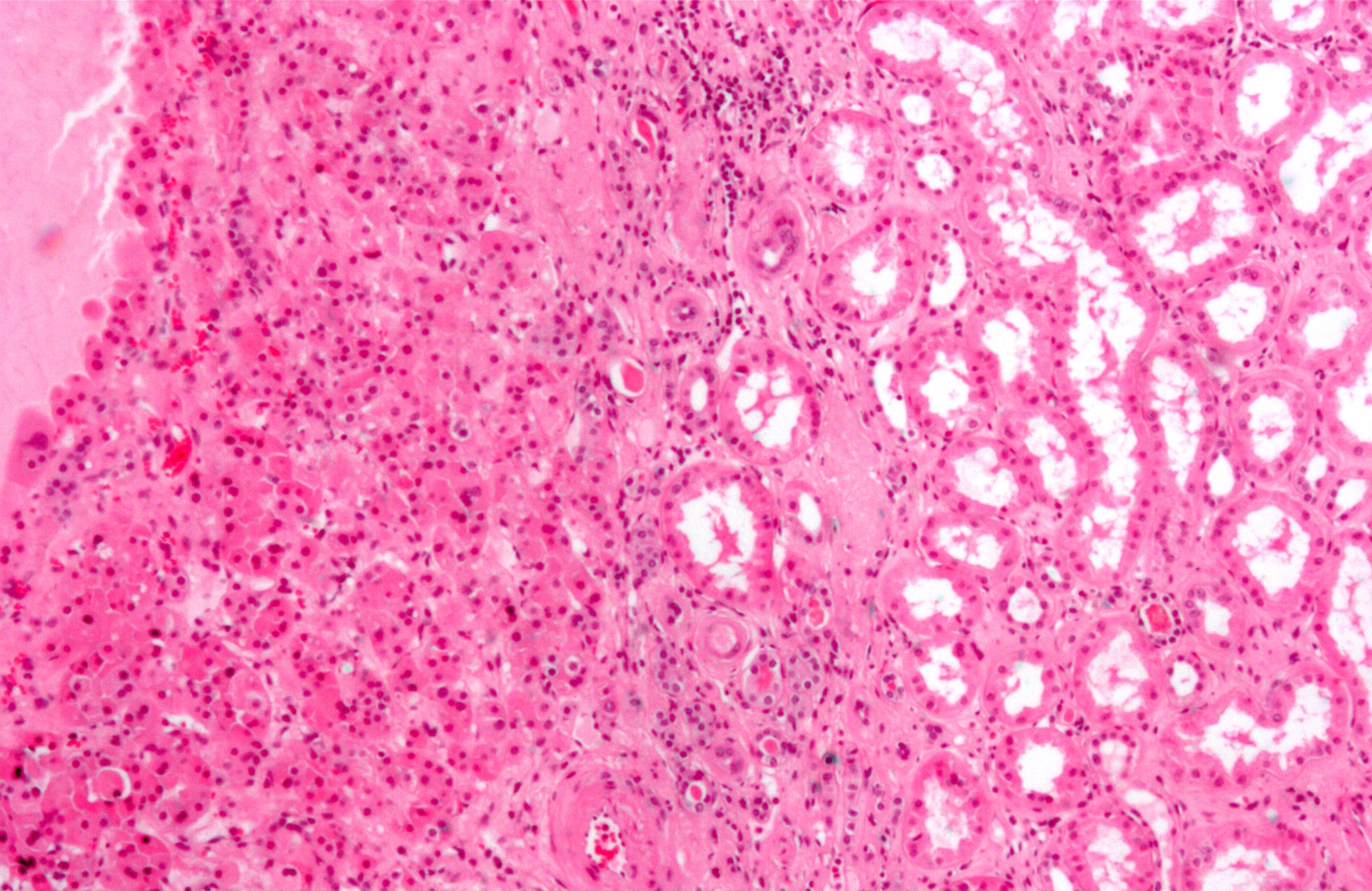
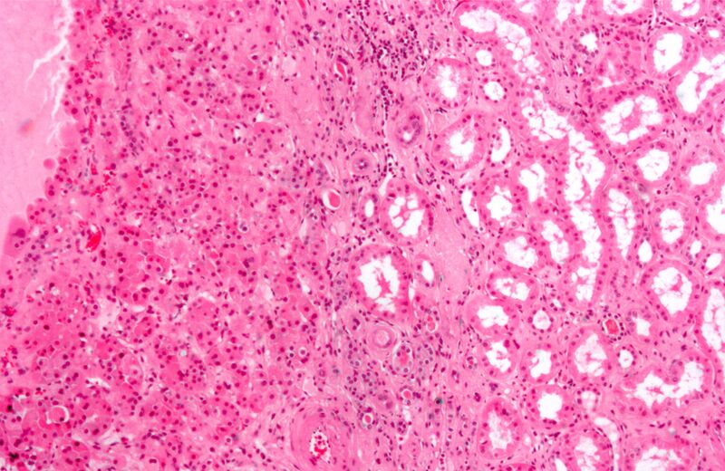
| Description | High magnification micrograph of a renal oncocytoma. H&E stain.
The tumour cells (left of the image) are arranged in nests, have slightly enlarged nuclei and have a more eosinophilic (darker pink) cytoplasm than the normal kidney - renal tubules (right of image). On electronmicroscopy, oncocytomas have abundant mitochondria. See also Image:Renal_oncocytoma3.jpg - intermediate magnification. Image:Renal_oncocytoma4.jpg - low magnification. |
| Source | Wikimedia Commons file page |
| Author | Nephron |
| Permission | See original Commons license details. |
Licensing[edit]
Creative Commons Attribution-ShareAlike 3.0 Unported (CC BY-SA 3.0)
This file is licensed under the Creative Commons Attribution-ShareAlike 3.0 license.
Official license: CC BY-SA 3.0
License page: CC BY-SA 3.0
Original attribution and file history: Wikimedia Commons
File history
Click on a date/time to view the file as it appeared at that time.
| Date/Time | Thumbnail | Dimensions | User | Comment | |
|---|---|---|---|---|---|
| current | 03:33, 5 June 2026 | | 1,952 × 1,268 (758 KB) | Maintenance script (talk | contribs) | == Summary == Importing file |
You cannot overwrite this file.
File usage
The following 3 pages use this file:
