File:Retroverted uterus in pregnancy.png
From WikiMD's WELLNESSPEDIA
No higher resolution available.
Retroverted_uterus_in_pregnancy.png (482 × 362 pixels, file size: 151 KB, MIME type: image/png)
Summary[edit]
| Summary | |
|---|---|
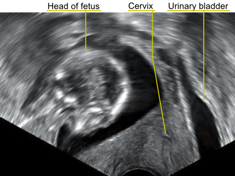
| Description | A transvaginal ultrasonography showing a retroverted uterus during a pregnancy of a gestational age of 16 weeks. The cervix lies posteriorly to the urinary bladder, and the uterus normally extends superiorly from it, but the direction of the body of the fetus reveals that the uterus extends backwards. |
| Source | Wikimedia Commons file page |
| Author | Mikael Häggström.
When using this image in external works, it may be cited as: Häggström, Mikael (2014). "Medical gallery of Mikael Häggström 2014". WikiJournal of Medicine 1 (2). DOI:10.15347/wjm/2014.008. ISSN 2002-4436. Public Domain. or By Mikael Häggström, used with permission. |
| Permission | See original Commons license details. |
Licensing[edit]
License: CC0
License page: CC0
Original attribution and file history: Wikimedia Commons
File history
Click on a date/time to view the file as it appeared at that time.
| Date/Time | Thumbnail | Dimensions | User | Comment | |
|---|---|---|---|---|---|
| current | 22:28, 8 June 2026 | | 482 × 362 (151 KB) | Maintenance script (talk | contribs) | == Summary == Importing file |
You cannot overwrite this file.
File usage
The following page uses this file:
