File:Rhodopsin-transducin.png
From WikiMD's WELLNESSPEDIA
Size of this preview: 483 × 599 pixels. Other resolution: 1,516 × 1,880 pixels.
Original file (1,516 × 1,880 pixels, file size: 1.52 MB, MIME type: image/png)
Summary[edit]
| Summary | |
|---|---|
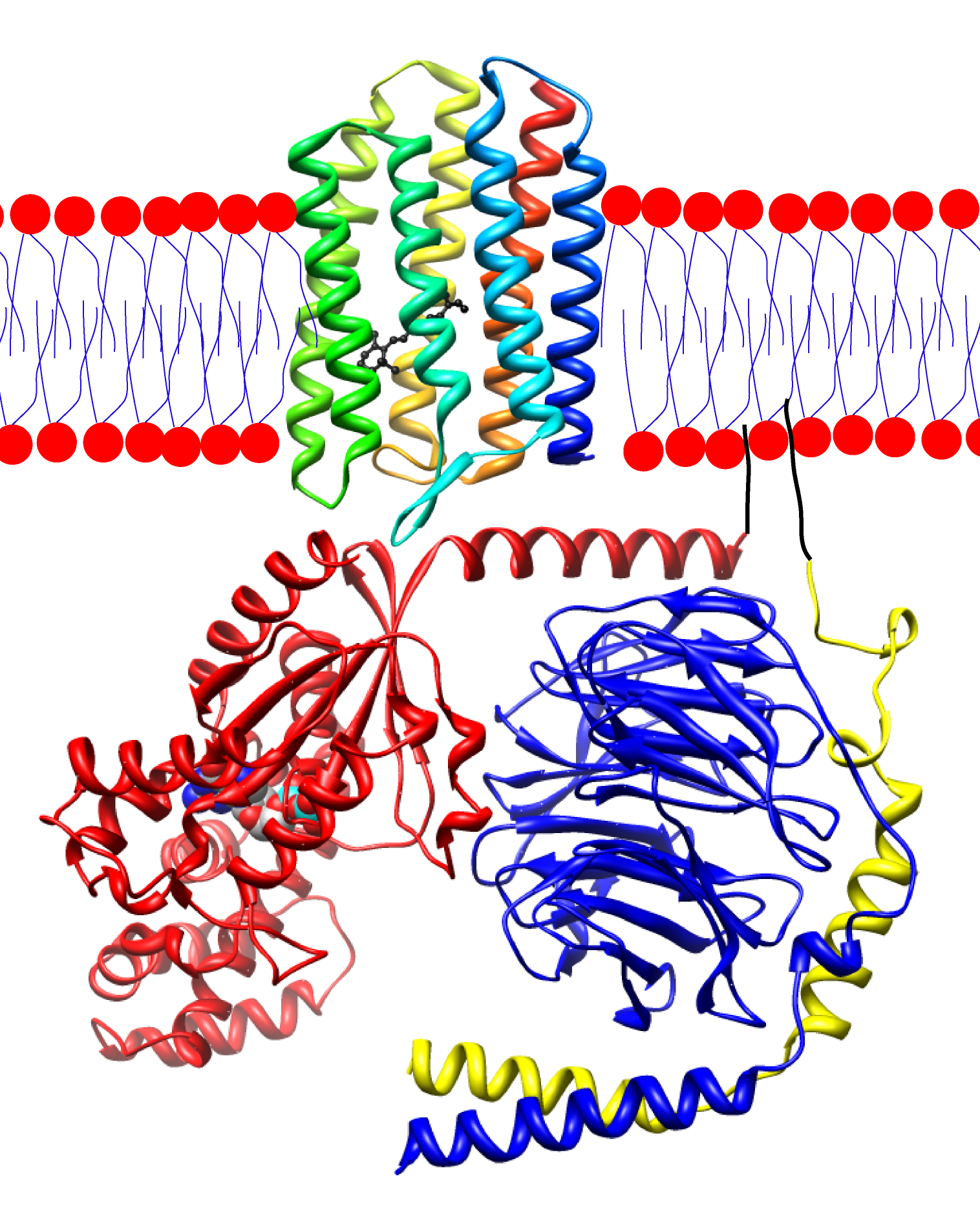
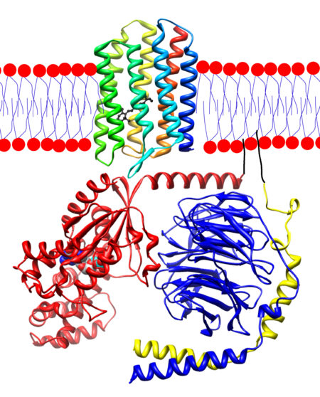
| Description | Sensory rhodopsin II (1gue.pdb) embedded in the membrane with transducin (1got.pdb) under it. Rhodopsin is colored in a rainbow with the N-terminus red and the C-terminus blue. There is a bound retinal on the inside that I've colored black for ease of visualization. For the transducin, the Gt-alpha subunit is red, beta is blue and gamma is yellow. I've drawn in pseudo anchoring sites in black. The Gt-alpha subunit has a bound GDP that is colored by atom. The protein structures were created using UCSF chimera and then placed together in adobe illustrator. This illustration is entirely my own using the publicly available pdb data. I'm releasing it into the public domain since I couldn't find any nice illustrations of a rhodopsin and transducin. |
| Source | Wikimedia Commons |
| Author | Dpryan at English Wikipedia |
| Permission | See Commons |
Licensing[edit]
Public Domain
This file is in the public domain and may be used without restriction.
Please see the linked source page for the original file history, attribution information, and licensing details.
Original attribution and file history: Wikimedia Commons
File history
Click on a date/time to view the file as it appeared at that time.
| Date/Time | Thumbnail | Dimensions | User | Comment | |
|---|---|---|---|---|---|
| current | 01:22, 2 June 2026 | | 1,516 × 1,880 (1.52 MB) | Maintenance script (talk | contribs) | == Summary == Importing file |
You cannot overwrite this file.
File usage
The following file is a duplicate of this file (more details):
- File:Rhodopsin-transducin.png from Wikimedia Commons
The following 2 pages use this file:
