File:Seminoma high mag.jpg
From WikiMD's WELLNESSPEDIA
Size of this preview: 800 × 533 pixels. Other resolutions: 2,560 × 1,707 pixels | 4,272 × 2,848 pixels.
Original file (4,272 × 2,848 pixels, file size: 4.83 MB, MIME type: image/jpeg)
Summary[edit]
| Summary | |
|---|---|
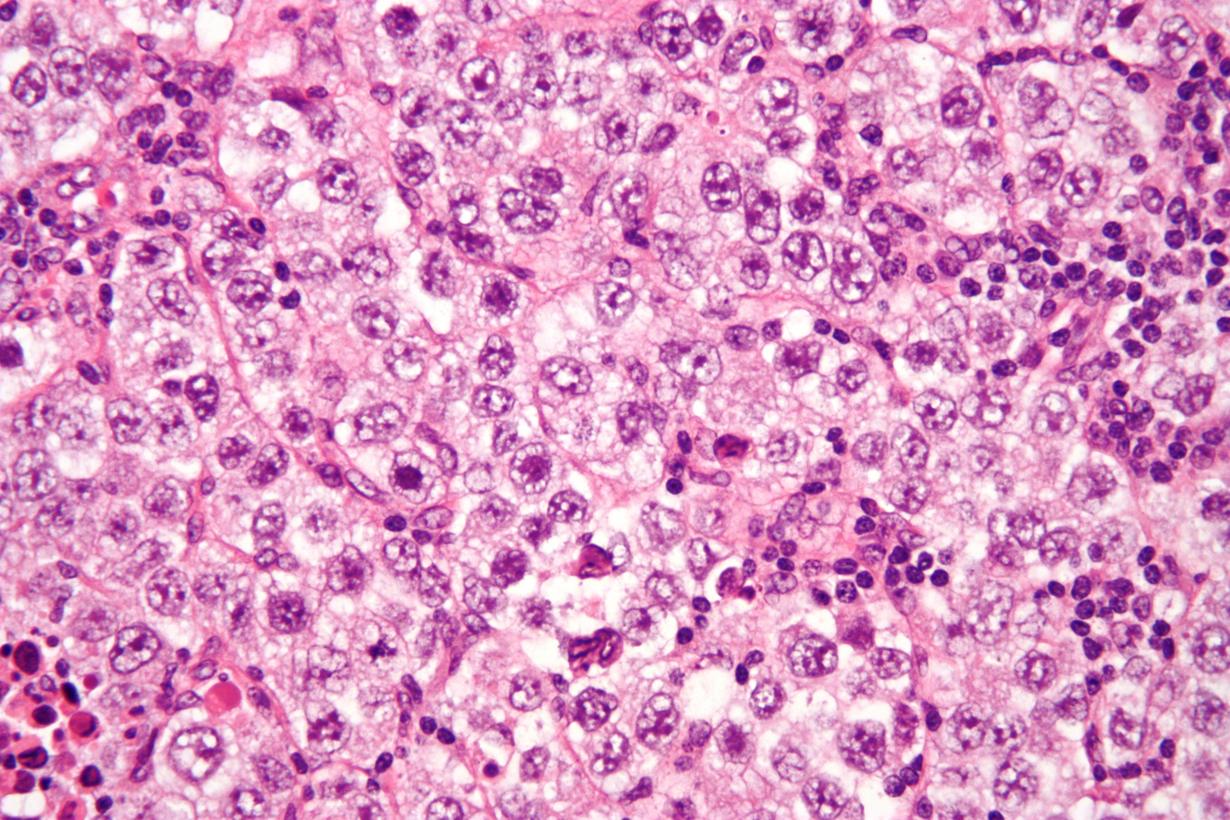
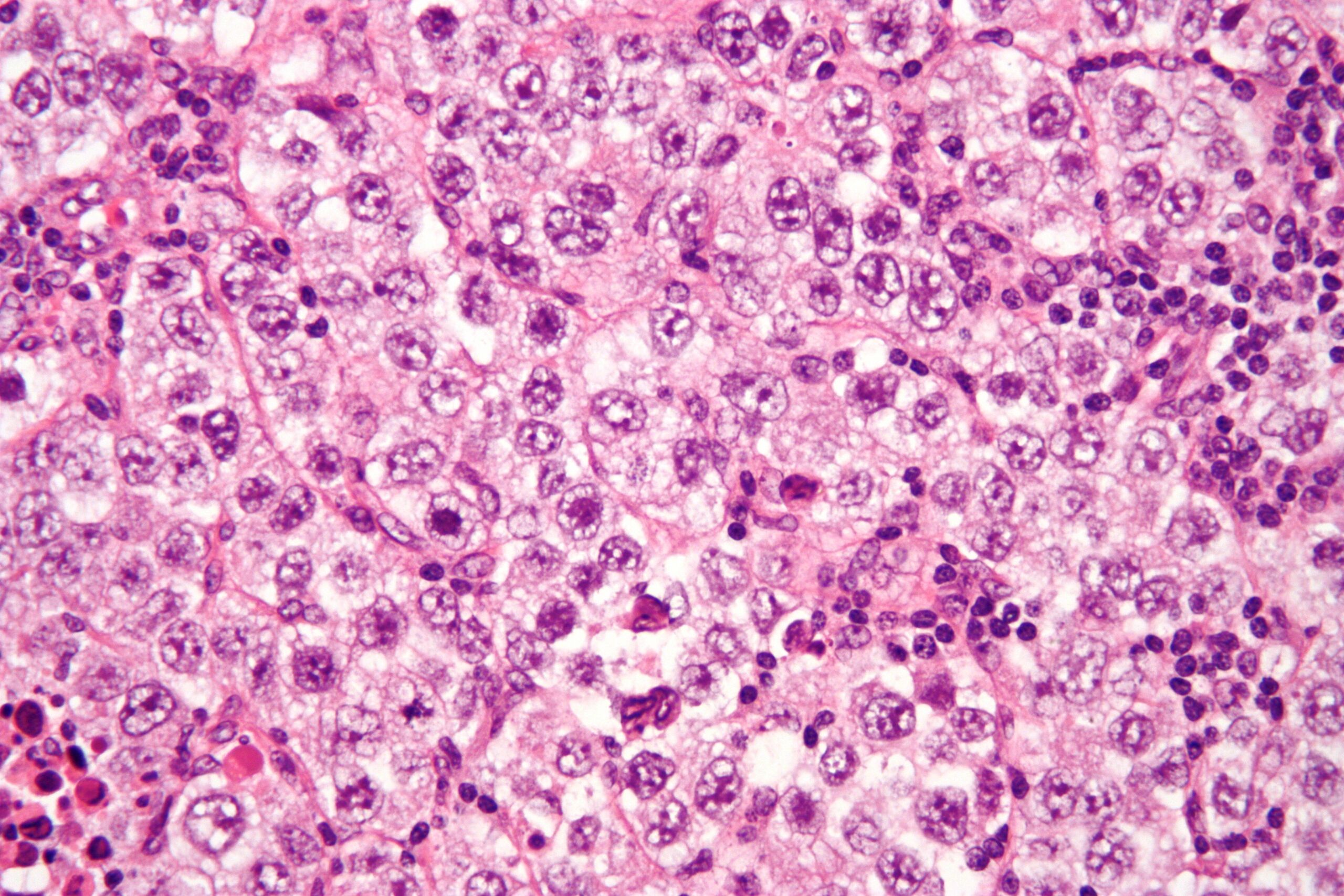
| Description | Micrograph of a seminoma. H&E stain.
Features: Lymphocytes. Tumour cells with a fried egg-like appearance: Central nucleus with nucleolus. Clear cytoplasm. Well-defined cell borders. Related images
Intermed. mag.
High mag.
|
| Source | Wikimedia Commons |
| Author | Nephron |
| Permission | See Commons |
Licensing[edit]
Creative Commons Attribution-ShareAlike 3.0 Unported (CC BY-SA 3.0)
This file is licensed under the Creative Commons Attribution-ShareAlike 3.0 license.
Official license: CC BY-SA 3.0
Original attribution and file history: Wikimedia Commons
File history
Click on a date/time to view the file as it appeared at that time.
| Date/Time | Thumbnail | Dimensions | User | Comment | |
|---|---|---|---|---|---|
| current | 03:41, 2 June 2026 | | 4,272 × 2,848 (4.83 MB) | Maintenance script (talk | contribs) | == Summary == Importing file |
You cannot overwrite this file.
File usage
The following 2 pages use this file:
