File:Serous carcinoma cytology.jpg

Original file (4,272 × 2,848 pixels, file size: 870 KB, MIME type: image/jpeg)
Summary[edit]
| Summary | |
|---|---|
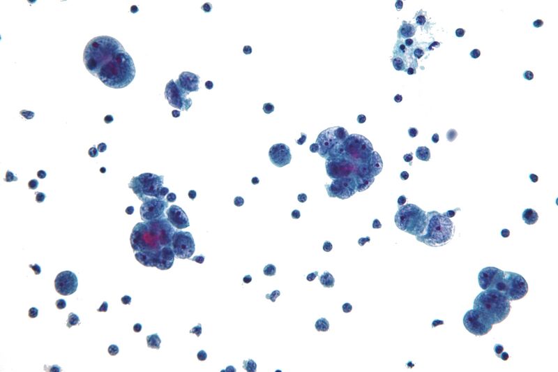
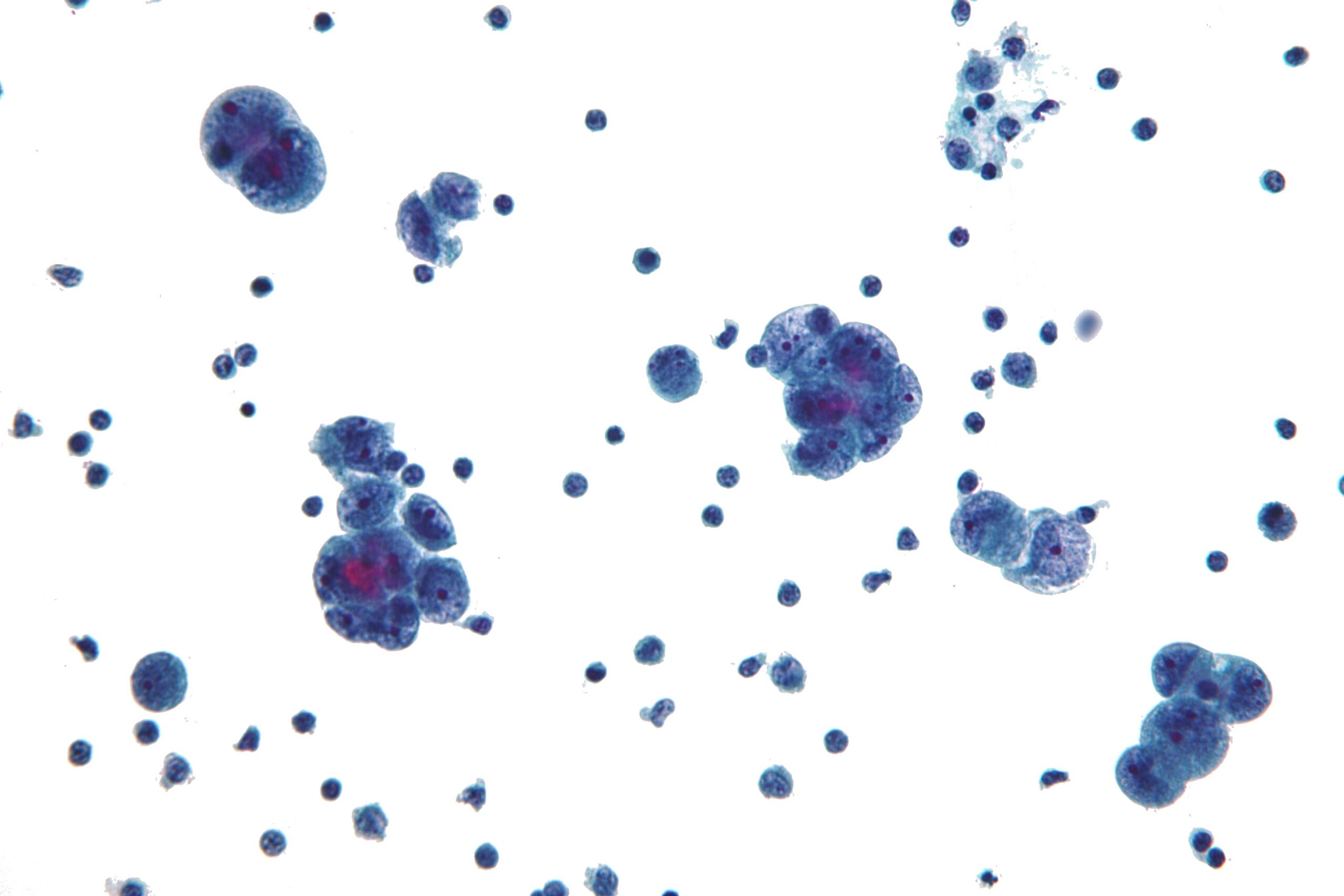
| Description | Micrograph showing serous carcinoma. Peritoneal fluid specimen.
Features: Marked intragroup nuclear pleomorphism. Macronucleoli. "Knobby" group borders (in large groups) - not apparent on image. Hydropic vacuoles - not apparent on image. Differential diagnosis of serous carcinoma: Ovarian serous carcinoma. Uterine serous carcinoma. Fallopian tube serous carcinoma. Cervix serous carcinoma. Primary peritoneal serous carcinoma. Related images
Animation showing 3-D nature of cluster.
Component of animation.
Component of animation.
Another case.
Low mag.
|
| Source | Wikimedia Commons file page |
| Author | Nephron |
| Permission | See original Commons license details. |
Licensing[edit]
Creative Commons Attribution-ShareAlike 3.0 Unported (CC BY-SA 3.0)
This file is licensed under the Creative Commons Attribution-ShareAlike 3.0 license.
Official license: CC BY-SA 3.0
License page: CC BY-SA 3.0
Original attribution and file history: Wikimedia Commons
File history
Click on a date/time to view the file as it appeared at that time.
| Date/Time | Thumbnail | Dimensions | User | Comment | |
|---|---|---|---|---|---|
| current | 03:34, 5 June 2026 | | 4,272 × 2,848 (870 KB) | Maintenance script (talk | contribs) | == Summary == Importing file |
You cannot overwrite this file.
File usage
The following file is a duplicate of this file (more details):
- File:Serous carcinoma cytology.jpg from Wikimedia Commons
The following 4 pages use this file:
