File:Sinoatrial node 2 low mag.jpg
Original file (2,588 × 2,054 pixels, file size: 2.32 MB, MIME type: image/jpeg)
Summary[edit]
| Summary | |
|---|---|
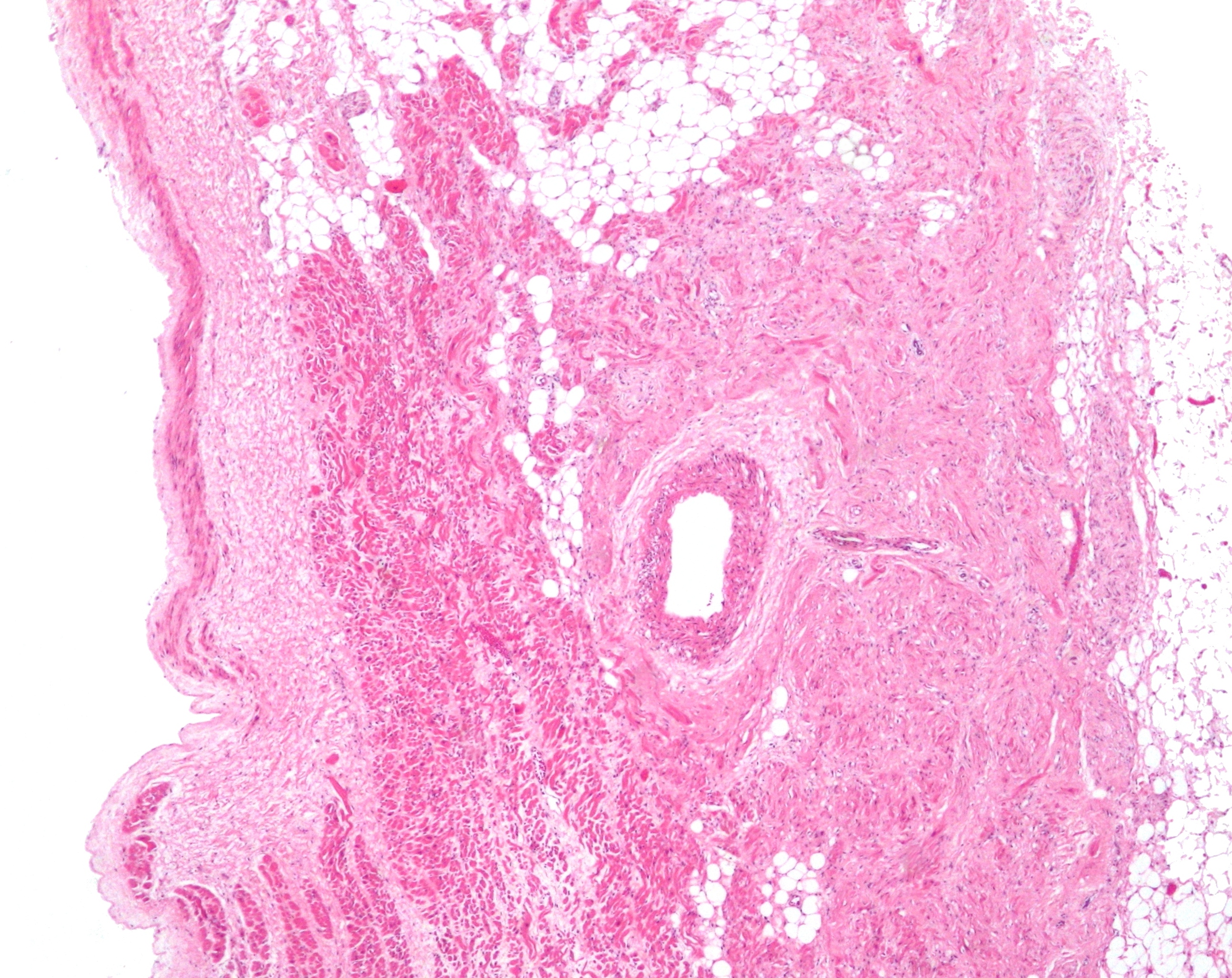
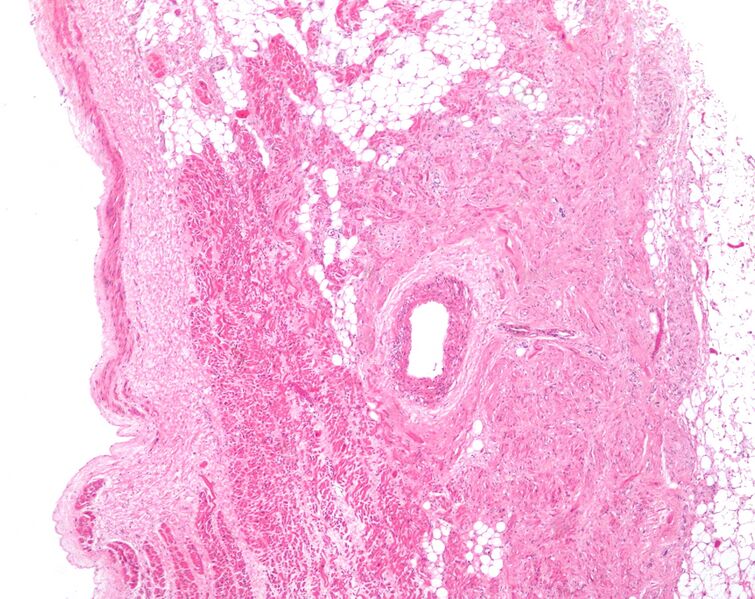
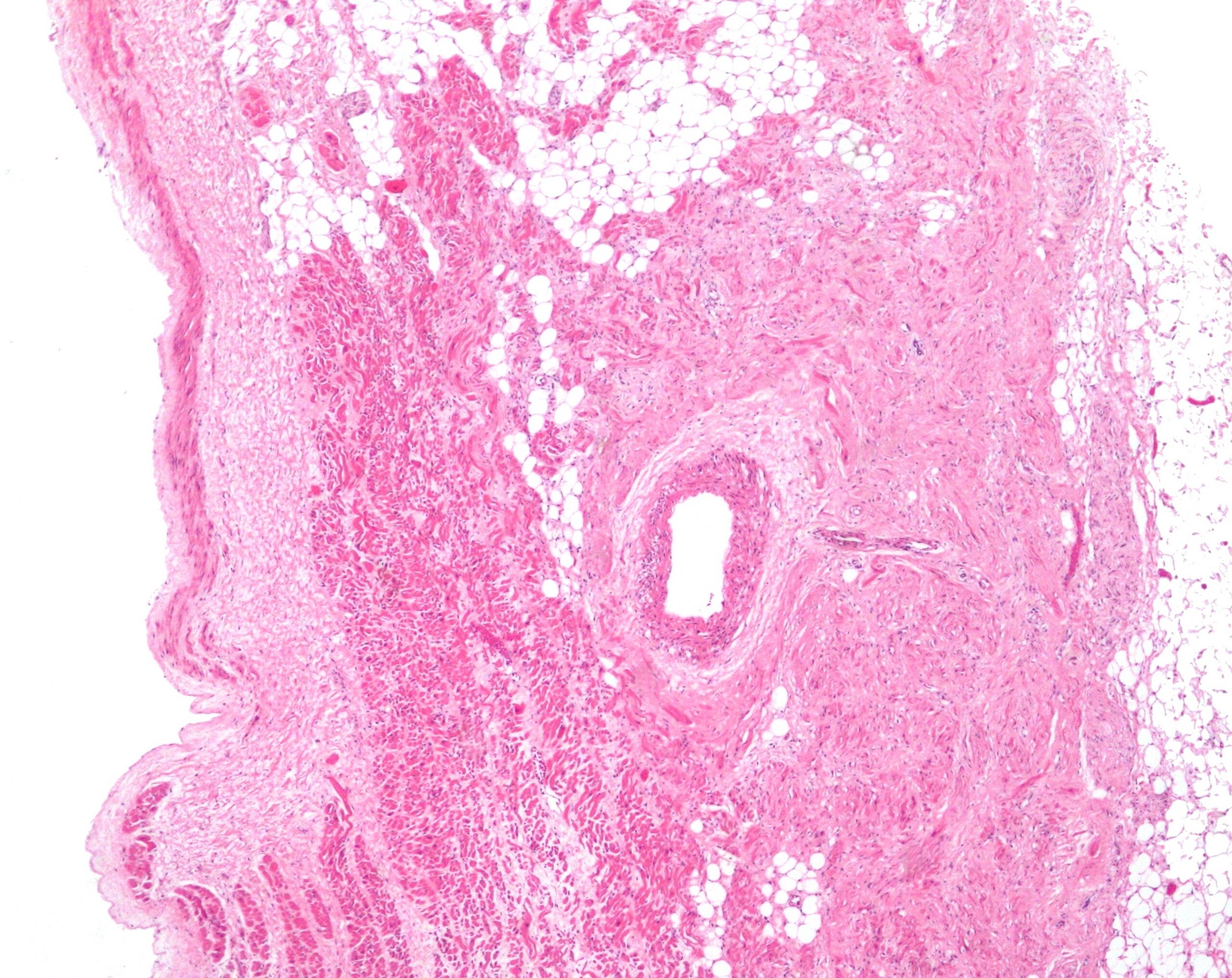
| Description | Micrograph of the sinoatrial (SA) node. H&E stain.
The SA node fibre vaguely resemble cardiac myocytes; however, they are thinner, squiggly and stain less intensely (on H&E) than cardiac myocytes. Description of the micrograph - the follow things are seen: Cardiac myocytes of the right atrium are seen at the right/bottom. Nodal artery, a branch of the right coronary artery is seen in the centre - on lumen. Sinoatrial node surrounds the nodal artery and abuts the darker staining cardiac myocytes. Lumen of the right atrium is on the left (endothelial lining not seen). Epicardial apidose tissue is to the right of the SA node. Epicardial space is on the right of the image. Adjacent to nodal tissue is a nerve; the SA node interacts with fibres from the vagus nerve. Related images
High mag. image of the same SA node.
|
| Source | Wikimedia Commons |
| Author | Nephron |
| Permission | See Commons |
Licensing[edit]
Creative Commons Attribution-ShareAlike 3.0 Unported (CC BY-SA 3.0)
This file is licensed under the Creative Commons Attribution-ShareAlike 3.0 license.
Official license: CC BY-SA 3.0
Original attribution and file history: Wikimedia Commons
File history
Click on a date/time to view the file as it appeared at that time.
| Date/Time | Thumbnail | Dimensions | User | Comment | |
|---|---|---|---|---|---|
| current | 03:40, 2 June 2026 | | 2,588 × 2,054 (2.32 MB) | Maintenance script (talk | contribs) | == Summary == Importing file |
You cannot overwrite this file.
File usage
The following file is a duplicate of this file (more details):
- File:Sinoatrial node 2 low mag.jpg from Wikimedia Commons
The following 2 pages use this file:
