File:Squamous carcinoma lung cytology.gif

Original file (2,136 × 1,424 pixels, file size: 1.54 MB, MIME type: image/gif, looped, 2 frames, 2.0 s)
Summary[edit]
| Summary | |
|---|---|
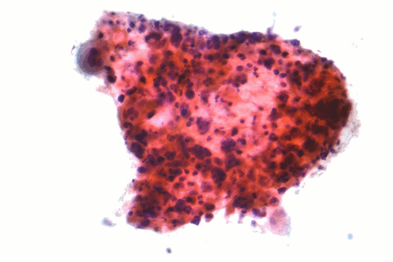
| Description | Micrographs of squamous carcinoma, also squamous cell carcinoma. FNA specimen of a lung lesion. Pap stain. Clinicoradiological findings in the case suggested it was a lung primary, i.e. lung cancer.
The micrographs show a 3-dimensional cluster of cells with the nuclear changes of malignancy (variation in nuclear size, staining and shape), and features of squamous differentiation (moderate amount of cytoplasm, small/indistinct nucleoli, nucleus in the cell centre, keratinization). See also Image:Small cell lung cancer - cytology.jpg |
| Source | Wikimedia Commons file page |
| Author | Nephron |
| Permission | See original Commons license details. |
Licensing[edit]
Creative Commons Attribution-ShareAlike 3.0 Unported (CC BY-SA 3.0)
This file is licensed under the Creative Commons Attribution-ShareAlike 3.0 license.
Official license: CC BY-SA 3.0
Original attribution and file history: Wikimedia Commons
File history
Click on a date/time to view the file as it appeared at that time.
| Date/Time | Thumbnail | Dimensions | User | Comment | |
|---|---|---|---|---|---|
| current | 12:51, 29 May 2026 | | 2,136 × 1,424 (1.54 MB) | Maintenance script (talk | contribs) | == Summary == Importing file |
You cannot overwrite this file.
File usage
The following file is a duplicate of this file (more details):
- File:Squamous carcinoma lung cytology.gif from Wikimedia Commons
The following 2 pages use this file:
