File:Tauopathy in Alzheimer's disease.jpg
From WikiMD's WELLNESSPEDIA
Size of this preview: 565 × 599 pixels. Other resolution: 2,400 × 2,546 pixels.
Original file (2,400 × 2,546 pixels, file size: 2.44 MB, MIME type: image/jpeg)
Summary[edit]
| Summary | |
|---|---|
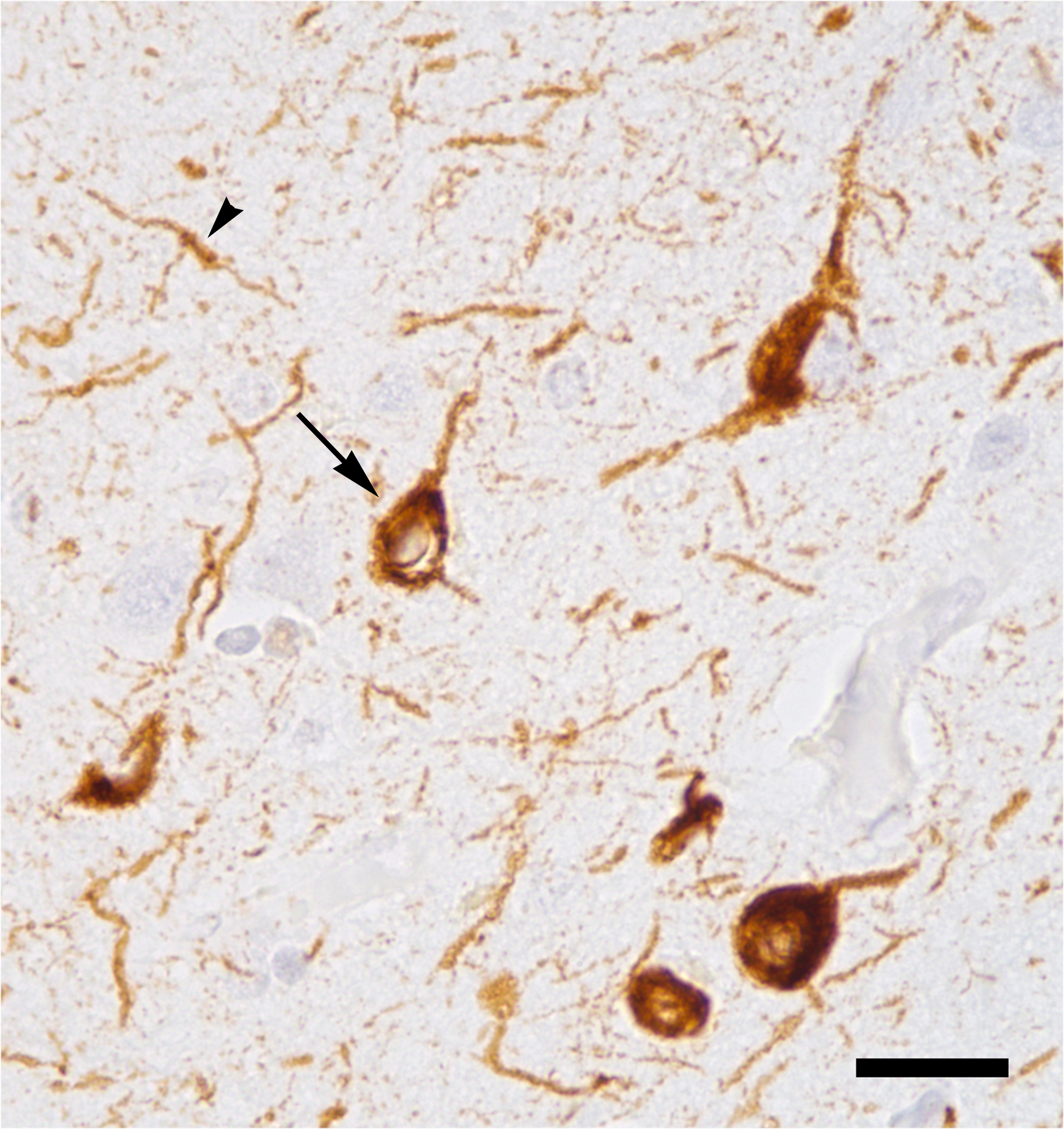
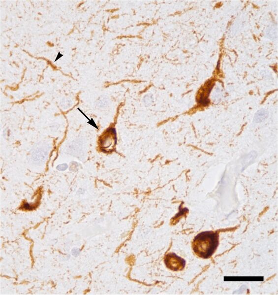
| Description | This photomicrograph shows nerve cell bodies (one is indicated by the arrow) and their processes (one is indicated by the arrowhead) in the neocortex of a patient who had died with Alzheimer's disease at Braak stage VI. Tau protein is stained brown using immunohistochemistry. The tissue section is lightly counterstained with hematoxylin (light blue). The bar represents a distance of 25 microns (0.025 millimeters). |
| Source | Wikimedia Commons file page |
| Author | Tulemo |
| Permission | See original Commons license details. |
Licensing[edit]
Creative Commons Attribution-ShareAlike 4.0 International (CC BY-SA 4.0)
This file is licensed under the Creative Commons Attribution-ShareAlike 4.0 International license.
You are free to:
- Share — copy and redistribute the material.
- Adapt — remix, transform, and build upon the material.
Under the following conditions:
- Attribution — appropriate credit must be given.
- ShareAlike — derivative works must be distributed under the same license.
Official license: CC BY-SA 4.0
License page: CC BY-SA 4.0
Original attribution and file history: Wikimedia Commons
File history
Click on a date/time to view the file as it appeared at that time.
| Date/Time | Thumbnail | Dimensions | User | Comment | |
|---|---|---|---|---|---|
| current | 22:37, 8 June 2026 | | 2,400 × 2,546 (2.44 MB) | Maintenance script (talk | contribs) | == Summary == Importing file |
You cannot overwrite this file.
File usage
The following file is a duplicate of this file (more details):
- File:Tauopathy in Alzheimer's disease.jpg from Wikimedia Commons
The following page uses this file:
