File:Telocytes - Fig 1.jpg
From WikiMD's WELLNESSPEDIA
Size of this preview: 800 × 255 pixels. Other resolution: 5,317 × 1,692 pixels.
Original file (5,317 × 1,692 pixels, file size: 6.02 MB, MIME type: image/jpeg)
Summary[edit]
| Summary | |
|---|---|
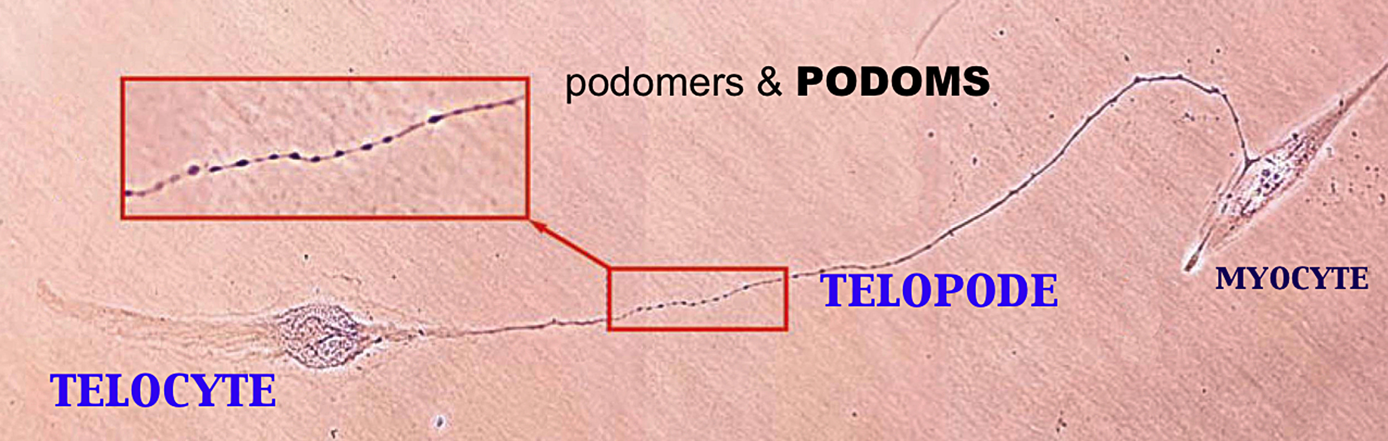
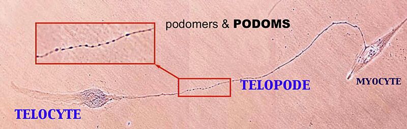
| Description | Human non-pregnant myometrium in cell culture; day 3; the first passage. Giemsa staining. One TC establishing contacts with a myocyte by a Tp of about 65 mm long. Photographic composition of 4 serial phase contrast images; original magnification 40x. In red rectangles, a higher magnification clearly shows the moniliform aspect. At least 40 specific dilations (podoms) interconnected by thin segments (podomeres) are visible in a ‘beadlike’ fashion. |
| Source | Wikimedia Commons file page |
| Author | Lmpopescu |
| Permission | See original Commons license details. |
Licensing[edit]
Creative Commons Attribution-ShareAlike 3.0 Unported (CC BY-SA 3.0)
This file is licensed under the Creative Commons Attribution-ShareAlike 3.0 license.
Official license: CC BY-SA 3.0
License page: CC BY-SA 3.0
Original attribution and file history: Wikimedia Commons
File history
Click on a date/time to view the file as it appeared at that time.
| Date/Time | Thumbnail | Dimensions | User | Comment | |
|---|---|---|---|---|---|
| current | 22:30, 8 June 2026 | 5,317 × 1,692 (6.02 MB) | Maintenance script (talk | contribs) | == Summary == Importing file |
You cannot overwrite this file.
File usage
The following file is a duplicate of this file (more details):
- File:Telocytes - Fig 1.jpg from Wikimedia Commons
The following page uses this file:
