File:Telocytes - Fig 2.jpg
From WikiMD's WELLNESSPEDIA

Size of this preview: 800 × 458 pixels. Other resolution: 1,947 × 1,114 pixels.
Original file (1,947 × 1,114 pixels, file size: 2 MB, MIME type: image/jpeg)
Summary[edit]
| Summary | |
|---|---|
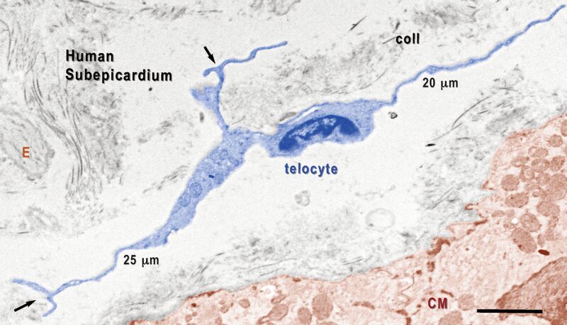
| Description | Digitally coloured TEM image shows TC (blue) in human subepicardium, bordering the peripheral cardiomyocytes (CM, highlighted in brown). The TC has three telopodes, illustrating: a) the distinctive dichotomous pattern of branching (arrows); b) Tp are very thin at the emergence of the cell body; c) alternating podoms and podomeres. Note that some portions of podomeres have the same thickness as collagen fibrills, which make them impossible to be observed under light microscopy. E - elastin Scale bar - 2 mm. |
| Source | Wikimedia Commons file page |
| Author | Lmpopescu |
| Permission | See original Commons license details. |
Licensing[edit]
Creative Commons Attribution-ShareAlike 3.0 Unported (CC BY-SA 3.0)
This file is licensed under the Creative Commons Attribution-ShareAlike 3.0 license.
Official license: CC BY-SA 3.0
License page: CC BY-SA 3.0
Original attribution and file history: Wikimedia Commons
File history
Click on a date/time to view the file as it appeared at that time.
| Date/Time | Thumbnail | Dimensions | User | Comment | |
|---|---|---|---|---|---|
| current | 22:31, 8 June 2026 | | 1,947 × 1,114 (2 MB) | Maintenance script (talk | contribs) | == Summary == Importing file |
You cannot overwrite this file.
File usage
The following page uses this file:
