File:Urethral urothelial cell carcinoma.jpg
Original file (4,272 × 2,848 pixels, file size: 3.58 MB, MIME type: image/jpeg)
Summary[edit]
| Summary | |
|---|---|
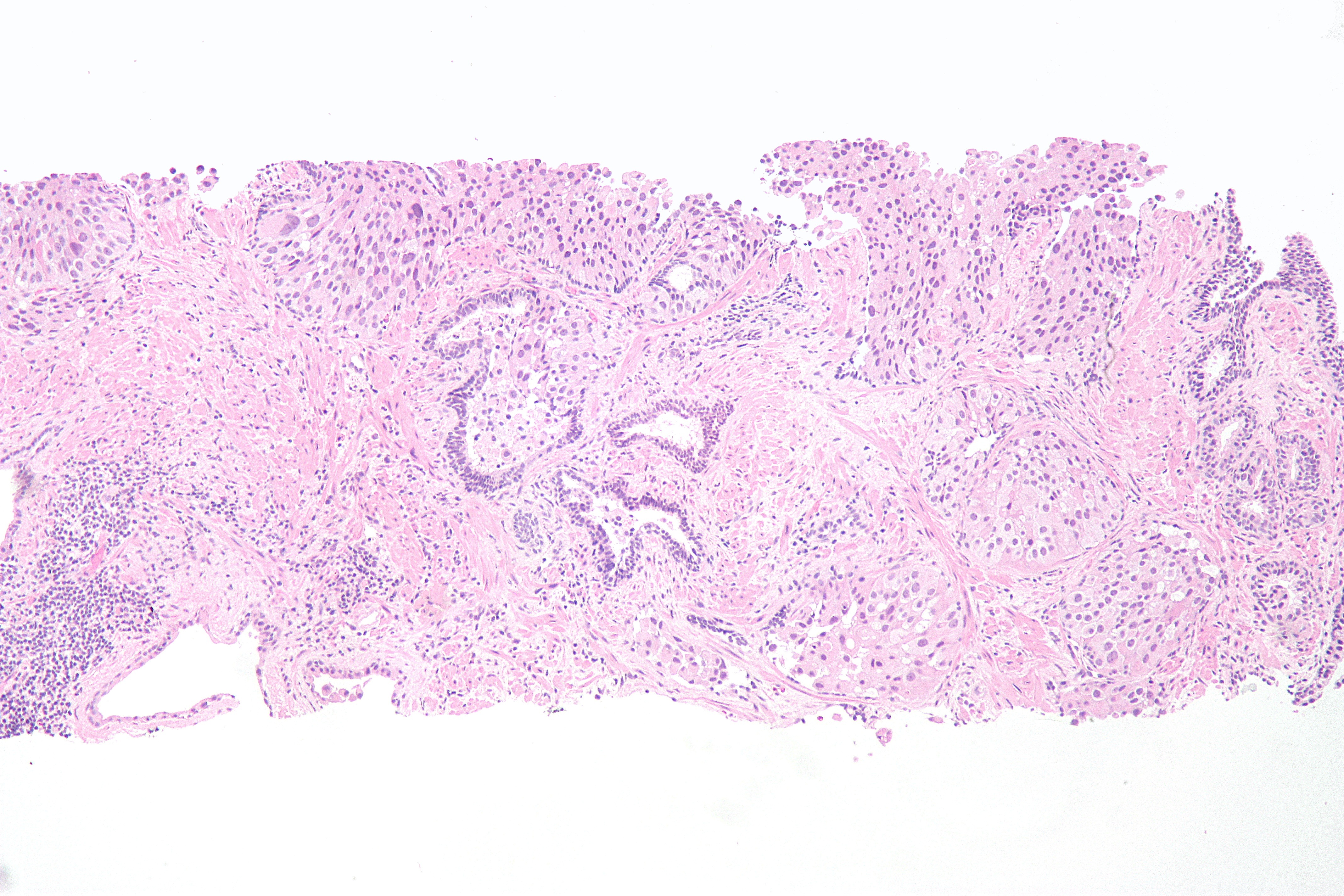
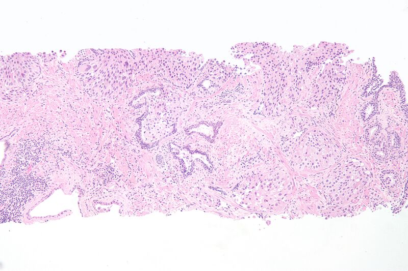
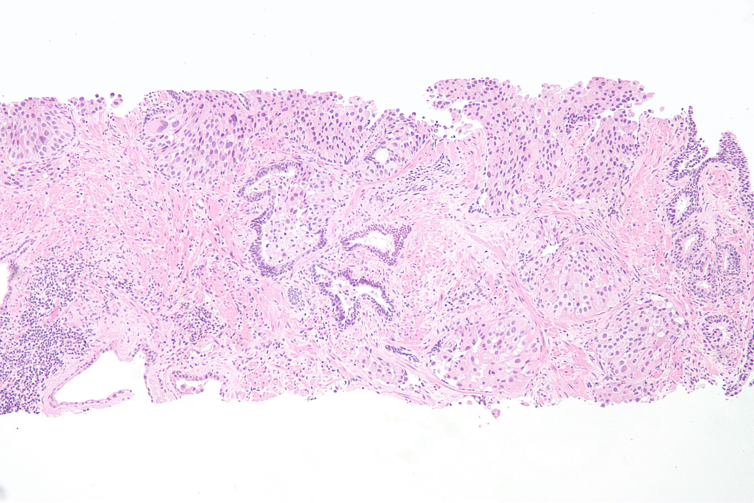
| Description | Micrograph of urothelial cell carcinoma of the prostatic urethra on a prostate biopsy. H&E stain.
Normal prostatic glands (with two cell layers) are seen on the far right of the image. At the top and centre of the image are nests of urothelial cell carcinoma, characterized by prominent nucleoli and marked variation of nuclear size. Slightly left-of-centre, it is possible to discern two cell populations in a prostatic gland: non-malignant prostatic epithelium with small darker stained nuclei, and neoplastic urothelium, with larger nuclei. |
| Source | Wikimedia Commons file page |
| Author | Nephron |
| Permission | See original Commons license details. |
Licensing[edit]
Creative Commons Attribution-ShareAlike 3.0 Unported (CC BY-SA 3.0)
This file is licensed under the Creative Commons Attribution-ShareAlike 3.0 license.
Official license: CC BY-SA 3.0
License page: CC BY-SA 3.0
Original attribution and file history: Wikimedia Commons
File history
Click on a date/time to view the file as it appeared at that time.
| Date/Time | Thumbnail | Dimensions | User | Comment | |
|---|---|---|---|---|---|
| current | 22:32, 8 June 2026 | | 4,272 × 2,848 (3.58 MB) | Maintenance script (talk | contribs) | == Summary == Importing file |
You cannot overwrite this file.
File usage
The following file is a duplicate of this file (more details):
- File:Urethral urothelial cell carcinoma.jpg from Wikimedia Commons
The following page uses this file:
