File:177-Apoptosomes apoptosomes.png
From WikiMD's WELLNESSPEDIA

Size of this preview: 800 × 501 pixels. Other resolution: 2,616 × 1,638 pixels.
Original file (2,616 × 1,638 pixels, file size: 2.69 MB, MIME type: image/png)
Summary[edit]
| Summary | |
|---|---|
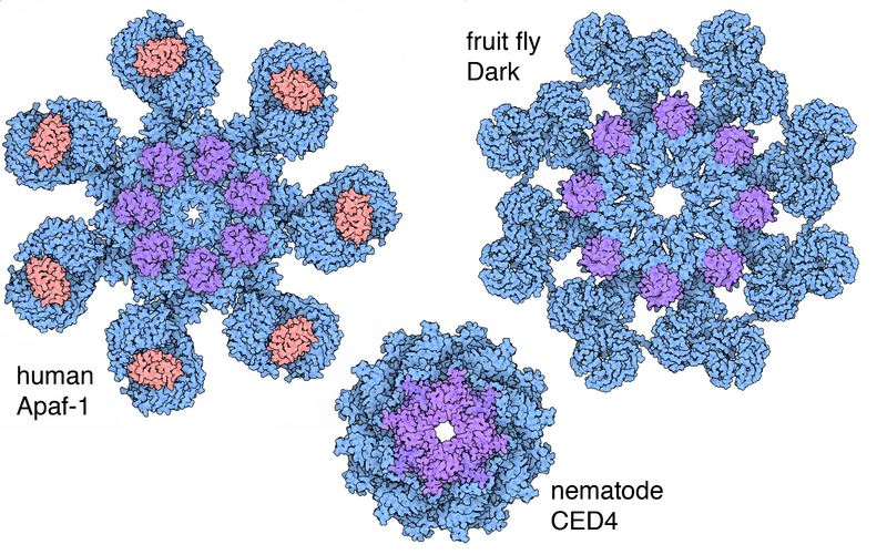
| Description | Space-fill drawing comparing the structures of the apoptosome molecular assemblies from human (Apaf-1), fruit fly (Dark), and nematode (CED4). The apoptosome subunits (shown in blue and purple) assemble when needed, to communicate between the system that signals cell death, such as cytochrome C for humans (in orange), to the system that disassembles the cell, such as the caspase proteases for humans, which are activated by binding to the CARD domains of the apoptosome (in purple). Image drawn by David Goodsell, from PDB files 3J2T, 3IZ8, and 3LQQ. |
| Source | Wikimedia Commons file page |
| Author | David Goodsell |
| Permission | See original Commons license details. |
Licensing[edit]
Creative Commons Attribution 3.0 Unported (CC BY 3.0)
This file is licensed under the Creative Commons Attribution 3.0 license.
Official license: CC BY 3.0
License page: CC BY 3.0
Original attribution and file history: Wikimedia Commons
File history
Click on a date/time to view the file as it appeared at that time.
| Date/Time | Thumbnail | Dimensions | User | Comment | |
|---|---|---|---|---|---|
| current | 03:22, 5 June 2026 | | 2,616 × 1,638 (2.69 MB) | Maintenance script (talk | contribs) | == Summary == Importing file |
You cannot overwrite this file.
File usage
The following file is a duplicate of this file (more details):
- File:177-Apoptosomes apoptosomes.png from Wikimedia Commons
The following 2 pages use this file:
