Muscle spindle
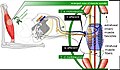
A Muscle spindle is a sensory receptor located in the muscle that senses changes in muscle length and velocity. These are also known as stretch receptors. They convey length information to the central nervous system via sensory neurons. This information can be processed by the brain to determine the position of body parts. The responses of muscle spindles to changes in length also play a crucial role in regulating the contraction of muscles, by supplying the motor system with the sensory feedback necessary to make fine adjustments to muscle force and position.
Structure[edit]
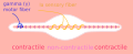
Muscle spindles are found within the belly of muscles, between extrafusal muscle fibers. The muscle spindle has both sensory and motor components. The sensory component consists of up to a dozen intrafusal muscle fibers, which are of two types: nuclear bag fibers and nuclear chain fibers. The motor component consists of gamma motor neurons.
Function[edit]
Muscle spindles have a variety of functions, including providing proprioceptive feedback to the central nervous system, contributing to fine motor control, and participating in the stretch reflex. When a muscle lengthens, the muscle spindle is stretched and its nerve activity increases. This increases alpha motor neuron activity, causing the muscle fibers to contract and thus resist the stretching. A secondary function is the measurement of the rate of change of muscle length, which can influence the long-term response of a muscle to a given length or velocity.
Clinical significance[edit]
Muscle spindles are involved in a number of clinical conditions, including spasticity, hypertonia, and Parkinson's disease. In these conditions, the sensitivity of the muscle spindle can be altered, leading to changes in muscle tone and movement.
See also[edit]
References[edit]

This WikiMD article can only be edited by registered and verified editors. You can log in or register.
- Muscle spindle
-
 Muscle spindle model
 -
 Muscle spindle light microscopy HE stain
 -
 Muscle spindle diagram
 -
 Muscle spindle
 -
 Muscle spindle
 -
 Muscle spindle
 -
 Fusimotor action


Medical Disclaimer: WikiMD is for informational purposes only and is not a substitute for professional medical advice. Content may be inaccurate or outdated and should not be used for diagnosis or treatment. Always consult your healthcare provider for medical decisions. Verify information with trusted sources such as CDC.gov and NIH.gov. By using this site, you agree that WikiMD is not liable for any outcomes related to its content. See full disclaimer.
Credits:Most images are courtesy of Wikimedia commons, and templates, categories Wikipedia, licensed under CC BY SA or similar.
Translate page: - East Asian
中文,
日本,
한국어,
South Asian
हिन्दी,
தமிழ்,
తెలుగు,
Urdu,
ಕನ್ನಡ,
Southeast Asian
Indonesian,
Vietnamese,
Thai,
မြန်မာဘာသာ,
বাংলা
European
español,
Deutsch,
français,
Greek,
português do Brasil,
polski,
română,
русский,
Nederlands,
norsk,
svenska,
suomi,
Italian
Middle Eastern & African
عربى,
Turkish,
Persian,
Hebrew,
Afrikaans,
isiZulu,
Kiswahili,
Other
Bulgarian,
Hungarian,
Czech,
Swedish,
മലയാളം,
मराठी,
ਪੰਜਾਬੀ,
ગુજરાતી,
Portuguese,
Ukrainian
