Schaeffer–Fulton stain
Schaeffer–Fulton stain is a differential staining technique used in microbiology to identify the presence of endospores in bacterial cells. This method distinguishes between the vegetative cells and the highly resistant endospores they can produce, which are significant in understanding bacterial life cycles, especially for those species capable of enduring extreme environments.
Principle[edit]
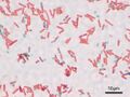
The Schaeffer–Fulton staining method utilizes two primary dyes: malachite green and safranin. Malachite green, the primary stain, is water-soluble and has a high affinity for the endospore coat, staining the endospores a bright green. Safranin is used as the counterstain, coloring the vegetative cells and any other cellular components pink or red. This contrast allows for the easy differentiation between the green-stained endospores and the red or pink-stained vegetative cells under a microscope.
Procedure[edit]
The staining process involves several steps:
- The bacterial smear is prepared on a slide and heat-fixed.
- Malachite green is applied to the slide, which is then steamed for several minutes to allow the dye to penetrate the endospores.
- The slide is rinsed with water, removing the malachite green from the vegetative cells but not the endospores.
- Safranin is applied as a counterstain, coloring the vegetative cells.
- The slide is then rinsed again, dried, and is ready for observation under a microscope.
Applications[edit]
Schaeffer–Fulton stain is widely used in microbiology for:
- Identifying bacterial species capable of forming endospores, which include genera such as Bacillus and Clostridium.
- Studying the life cycle of endospore-forming bacteria, particularly their response to environmental stress.
- Detecting the presence of endospores in clinical samples, soil, or food products, which is important in medical, environmental, and food microbiology.
Limitations[edit]
While the Schaeffer–Fulton stain is effective for endospore detection, it has limitations:
- It cannot differentiate between live and dead cells.
- The technique requires precise control of the staining and steaming process to avoid overstaining or understaining.
- It is specific to endospores and does not provide information on other cellular structures or functions.
See Also[edit]

This WikiMD article can only be edited by registered and verified editors. You can log in or register.
-
 Bacillus subtilis Spore


Medical Disclaimer: WikiMD is for informational purposes only and is not a substitute for professional medical advice. Content may be inaccurate or outdated and should not be used for diagnosis or treatment. Always consult your healthcare provider for medical decisions. Verify information with trusted sources such as CDC.gov and NIH.gov. By using this site, you agree that WikiMD is not liable for any outcomes related to its content. See full disclaimer.
Credits:Most images are courtesy of Wikimedia commons, and templates, categories Wikipedia, licensed under CC BY SA or similar.
Translate page: - East Asian
中文,
日本,
한국어,
South Asian
हिन्दी,
தமிழ்,
తెలుగు,
Urdu,
ಕನ್ನಡ,
Southeast Asian
Indonesian,
Vietnamese,
Thai,
မြန်မာဘာသာ,
বাংলা
European
español,
Deutsch,
français,
Greek,
português do Brasil,
polski,
română,
русский,
Nederlands,
norsk,
svenska,
suomi,
Italian
Middle Eastern & African
عربى,
Turkish,
Persian,
Hebrew,
Afrikaans,
isiZulu,
Kiswahili,
Other
Bulgarian,
Hungarian,
Czech,
Swedish,
മലയാളം,
मराठी,
ਪੰਜਾਬੀ,
ગુજરાતી,
Portuguese,
Ukrainian
