Gynecologic ultrasonography
Gynecologic ultrasonography or gynecologic sonography refers to the application of medical ultrasonography to the female pelvic organs, specifically the uterus, the ovaries, the Fallopian tubes, as well as the bladder, the adnexa, and the Pouch of Douglas. The procedure may lead to other medically relevant findings in the pelvis.
Indications[edit]
Gynecologic ultrasonography is used frequently in the diagnosis of many conditions. It can be used to investigate uterine problems such as menorrhagia, amenorrhea, abnormal vaginal bleeding and pain. It can also be used to identify ovarian cysts or masses, including ovarian cancer, ectopic pregnancy, and pelvic inflammatory disease.
Procedure[edit]
The procedure involves the use of a probe, which is inserted into the vagina after a protective cover is placed over it. The probe is moved within the vaginal cavity to scan the pelvic structures, while the patient lies on her back in a position similar to that used for a gynecologic exam.
Types[edit]
There are two main types of gynecologic ultrasonography: transvaginal and transabdominal. Transvaginal ultrasonography involves the insertion of a probe into the vagina, while transabdominal ultrasonography involves the application of a probe on the abdomen.
Risks[edit]
While gynecologic ultrasonography is generally considered safe, potential risks include discomfort during the transvaginal ultrasound and the possibility of an allergic reaction to the gel used during the transabdominal ultrasound.
See also[edit]

This WikiMD article can only be edited by registered and verified editors. You can log in or register.
-
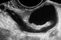 Hydrosalpinx (left)
 -
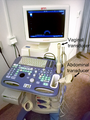 Transvaginal ultrasonography device
 -
 Mirena in optimal place - schematic
 -
Normal hysterosonography
-
Sonohysterography
.jpg)


Medical Disclaimer: WikiMD is for informational purposes only and is not a substitute for professional medical advice. Content may be inaccurate or outdated and should not be used for diagnosis or treatment. Always consult your healthcare provider for medical decisions. Verify information with trusted sources such as CDC.gov and NIH.gov. By using this site, you agree that WikiMD is not liable for any outcomes related to its content. See full disclaimer.
Credits:Most images are courtesy of Wikimedia commons, and templates, categories Wikipedia, licensed under CC BY SA or similar.
Translate page: - East Asian
中文,
日本,
한국어,
South Asian
हिन्दी,
தமிழ்,
తెలుగు,
Urdu,
ಕನ್ನಡ,
Southeast Asian
Indonesian,
Vietnamese,
Thai,
မြန်မာဘာသာ,
বাংলা
European
español,
Deutsch,
français,
Greek,
português do Brasil,
polski,
română,
русский,
Nederlands,
norsk,
svenska,
suomi,
Italian
Middle Eastern & African
عربى,
Turkish,
Persian,
Hebrew,
Afrikaans,
isiZulu,
Kiswahili,
Other
Bulgarian,
Hungarian,
Czech,
Swedish,
മലയാളം,
मराठी,
ਪੰਜਾਬੀ,
ગુજરાતી,
Portuguese,
Ukrainian
