Phonocardiogram
Phonocardiogram is a graphical recording of the sounds and murmurs made by the heart using the Phonocardiography technique. This non-invasive procedure is used to assess and diagnose various heart conditions.
Overview[edit]
A phonocardiogram (PCG) is a plot of high-frequency, low-amplitude sounds from the heart. These sounds are produced by the opening and closing of the heart's valves, and the flow of blood through the heart chambers. The PCG can provide valuable information about the timing of these events and the presence of any abnormal sounds or murmurs.
Procedure[edit]
The procedure for obtaining a phonocardiogram involves placing a special microphone on the chest, which is connected to an amplifier and a recording device. The patient is usually asked to lie still and breathe normally during the procedure. The sounds are then recorded and displayed on a graph.
Interpretation[edit]
The interpretation of a phonocardiogram involves correlating the recorded sounds with the phases of the Cardiac cycle. Normal heart sounds include the first heart sound (S1), produced by the closure of the mitral and tricuspid valves, and the second heart sound (S2), produced by the closure of the aortic and pulmonic valves. Additional sounds may indicate the presence of heart disease.
Clinical Significance[edit]
Phonocardiograms can be used to detect a variety of heart conditions, including Heart murmurs, Valvular heart disease, and Congenital heart disease. They can also be used to monitor the effectiveness of treatments for these conditions.
See Also[edit]
References[edit]

This WikiMD article can only be edited by registered and verified editors. You can log in or register.
-
Phonocardiogram
-
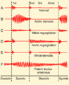 Phonocardiograms from normal and abnormal heart sounds
 -
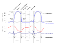 Wiggers Diagram
 -
William Birnbaum with a Phonocardiogram System


Medical Disclaimer: WikiMD is for informational purposes only and is not a substitute for professional medical advice. Content may be inaccurate or outdated and should not be used for diagnosis or treatment. Always consult your healthcare provider for medical decisions. Verify information with trusted sources such as CDC.gov and NIH.gov. By using this site, you agree that WikiMD is not liable for any outcomes related to its content. See full disclaimer.
Credits:Most images are courtesy of Wikimedia commons, and templates, categories Wikipedia, licensed under CC BY SA or similar.
Translate page: - East Asian
中文,
日本,
한국어,
South Asian
हिन्दी,
தமிழ்,
తెలుగు,
Urdu,
ಕನ್ನಡ,
Southeast Asian
Indonesian,
Vietnamese,
Thai,
မြန်မာဘာသာ,
বাংলা
European
español,
Deutsch,
français,
Greek,
português do Brasil,
polski,
română,
русский,
Nederlands,
norsk,
svenska,
suomi,
Italian
Middle Eastern & African
عربى,
Turkish,
Persian,
Hebrew,
Afrikaans,
isiZulu,
Kiswahili,
Other
Bulgarian,
Hungarian,
Czech,
Swedish,
മലയാളം,
मराठी,
ਪੰਜਾਬੀ,
ગુજરાતી,
Portuguese,
Ukrainian
