Posterior superior iliac spine

Anatomical landmark in the human pelvis
[[File: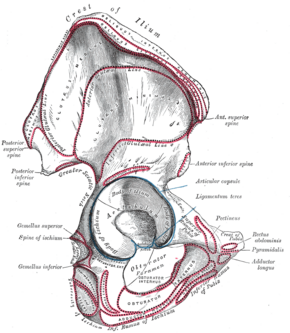 |frameless|alt=]]
| Details | |
|---|---|
| Synonyms | |
| Pronunciation | |
| Carnegie stage | |
| Days | |
| Precursor | Ilium (bone) |
| Gives rise to | |
| Part of | |
The posterior superior iliac spine (PSIS) is an important anatomical landmark located in the pelvis. It is a bony projection at the posterior end of the iliac crest of the ilium (bone), one of the three bones that make up the hip bone.
Anatomy[edit]
The PSIS is situated at the back of the pelvis and can be palpated through the skin. It is located at the level of the second sacral vertebra (S2) and serves as an attachment point for the posterior sacroiliac ligament and the thoracolumbar fascia. The PSIS is also a reference point for the sacroiliac joint, which connects the sacrum to the ilium.
Function[edit]
The PSIS plays a crucial role in the stability and movement of the pelvis. It serves as an attachment site for several important ligaments and muscles, including the erector spinae and the multifidus. These muscles are essential for maintaining posture and enabling movements such as bending and twisting.
Clinical Significance[edit]
The PSIS is often used as a landmark in various medical procedures, including lumbar punctures and epidural anesthesia. It is also a key reference point in the diagnosis and treatment of sacroiliac joint dysfunction and other lower back pain conditions. Palpation of the PSIS can help healthcare providers assess the alignment and symmetry of the pelvis.
Related Structures[edit]
See Also[edit]
References[edit]
This medical article is a stub. You can help WikiMD by expanding the page. |

Medical Disclaimer: WikiMD is for informational purposes only and is not a substitute for professional medical advice. Content may be inaccurate or outdated and should not be used for diagnosis or treatment. Always consult your healthcare provider for medical decisions. Verify information with trusted sources such as CDC.gov and NIH.gov. By using this site, you agree that WikiMD is not liable for any outcomes related to its content. See full disclaimer.
Credits:Most images are courtesy of Wikimedia commons, and templates, categories Wikipedia, licensed under CC BY SA or similar.
Translate page: - East Asian
中文,
日本,
한국어,
South Asian
हिन्दी,
தமிழ்,
తెలుగు,
Urdu,
ಕನ್ನಡ,
Southeast Asian
Indonesian,
Vietnamese,
Thai,
မြန်မာဘာသာ,
বাংলা
European
español,
Deutsch,
français,
Greek,
português do Brasil,
polski,
română,
русский,
Nederlands,
norsk,
svenska,
suomi,
Italian
Middle Eastern & African
عربى,
Turkish,
Persian,
Hebrew,
Afrikaans,
isiZulu,
Kiswahili,
Other
Bulgarian,
Hungarian,
Czech,
Swedish,
മലയാളം,
मराठी,
ਪੰਜਾਬੀ,
ગુજરાતી,
Portuguese,
Ukrainian
