The Trochlear Nerve
Anatomy > Gray's Anatomy of the Human Body > IX. Neurology > 5d. The Trochlear Nerve (N. Trochlearis; Fourth Nerve)
Henry Gray (1821–1865). Anatomy of the Human Body. 1918.
The Trochlear Nerve [edit]
(N. Trochlearis; Fourth Nerve)

The trochlear nerve (Fig. 776), the smallest of the cranial nerves, supplies the Obliquus superior oculi. It arises from a nucleus situated in the floor of the cerebral aqueduct, opposite the upper part of the inferior colliculus.
From its origin it runs downward through the tegmentum, and then turns backward into the upper part of the anterior medullary velum. Here it decussates with its fellow of the opposite side and emerges from the surface of the velum at the side of the frenulum veli, immediately behind the inferior colliculus.
The nerve is directed across the superior cerebellar peduncle, and then winds forward around the cerebral peduncle, immediately above the pons, pierces the dura mater in the free border of the tentorium cerebelli, just behind, and lateral to, the posterior clinoid process, and passes forward in the lateral wall of the cavernous sinus, between the oculomotor nerve and the ophthalmic division of the trigeminal.
It crosses the oculomotor nerve, and enters the orbit through the superior orbital fissure. It now becomes the highest of all the nerves, and lies medial to the frontal nerve. In the orbit it passes medialward, above the origin of the Levator palpebrae superioris, and finally enters the orbital surface of the Obliquus superior.
In the lateral wall of the cavernous sinus the trochlear nerve forms communications with the ophthalmic division of the trigeminal and with the cavernous plexus of the sympathetic.
In the superior orbital fissure it occasionally gives off a branch to the lacrimal nerve. It gives off a recurrent branch which passes backward between the layers of the tentorium cerebelli and divides into two or three filaments which may be traced as far as the wall of the transverse sinus.
Function[edit]
The trochlear nerve provides motor supply to the superior oblique muscle of the eye,The trochlear nerve carries axons of type GSE, general somatic efferent, which innervate skeletal muscle of the superior oblique muscle.
The superior oblique muscle ends in a tendon that passes through a fibrous loop, the trochlea, located anteriorly on the medial aspect of the orbit. Trochlea means “pulley” in Latin; the fourth nerve is named after this structure.
Additional images[edit]
-
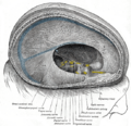 Dura mater and its processes exposed by removing part of the right half of the skull, and the brain.
 -
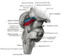 Hind- and mid-brains; postero-lateral view.
 -
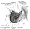 Dissection showing origins of right ocular muscles, and nerves entering by the superior orbital fissure.
 -
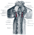 Upper part of medulla spinalis and hind- and mid-brains; posterior aspect, exposed in situ.
 -
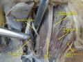 Trochlear nerve.Deep dissection.Superior view.


External links[edit]
- "Trochlear Nerve Palsy"
- lesson3 at The Anatomy Lesson by Wesley Norman (Georgetown University)
(orbit2
)
- cranialnerves at The Anatomy Lesson by Wesley Norman (Georgetown University)
(IV
)
- Animations of extraocular cranial nerve and muscle function and damage (University of Liverpool)
- Trochlear nerve at Neurolex
| The cranial nerves | ||||||||||
|---|---|---|---|---|---|---|---|---|---|---|
|
Gray's Anatomy[edit]
- Gray's Anatomy Contents
- Gray's Anatomy Subject Index
- About Classic Gray's Anatomy
- Glossary of anatomy terms
Anatomy atlases (external)[edit]
[1] - Anatomy Atlases
| Human systems and organs | ||||||||||||||
|---|---|---|---|---|---|---|---|---|---|---|---|---|---|---|
|

Medical Disclaimer: WikiMD is for informational purposes only and is not a substitute for professional medical advice. Content may be inaccurate or outdated and should not be used for diagnosis or treatment. Always consult your healthcare provider for medical decisions. Verify information with trusted sources such as CDC.gov and NIH.gov. By using this site, you agree that WikiMD is not liable for any outcomes related to its content. See full disclaimer.
Credits:Most images are courtesy of Wikimedia commons, and templates, categories Wikipedia, licensed under CC BY SA or similar.
Translate page: - East Asian
中文,
日本,
한국어,
South Asian
हिन्दी,
தமிழ்,
తెలుగు,
Urdu,
ಕನ್ನಡ,
Southeast Asian
Indonesian,
Vietnamese,
Thai,
မြန်မာဘာသာ,
বাংলা
European
español,
Deutsch,
français,
Greek,
português do Brasil,
polski,
română,
русский,
Nederlands,
norsk,
svenska,
suomi,
Italian
Middle Eastern & African
عربى,
Turkish,
Persian,
Hebrew,
Afrikaans,
isiZulu,
Kiswahili,
Other
Bulgarian,
Hungarian,
Czech,
Swedish,
മലയാളം,
मराठी,
ਪੰਜਾਬੀ,
ગુજરાતી,
Portuguese,
Ukrainian
