Outer enamel epithelium
Layer of cells in the enamel organ
[[File: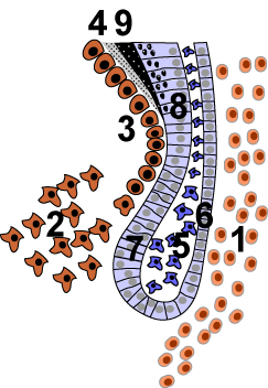 |frameless|alt=]]

| Details | |
|---|---|
| Synonyms | |
| Pronunciation | |
| Carnegie stage | |
| Days | |
| Precursor | |
| Gives rise to | |
| Part of | |
The outer enamel epithelium (OEE) is a layer of cells that forms part of the enamel organ during the development of a tooth. It plays a crucial role in the formation and protection of the developing enamel.
Structure[edit]
The outer enamel epithelium is composed of a single layer of cuboidal cells. These cells are located on the outer surface of the enamel organ, which is a key structure in the development of teeth. The OEE is continuous with the inner enamel epithelium at the cervical loop, a region critical for the growth and shaping of the tooth.
Function[edit]
The primary function of the outer enamel epithelium is to serve as a protective barrier for the developing enamel organ. It helps maintain the shape of the enamel organ and provides a boundary between the enamel organ and the surrounding dental follicle. Additionally, the OEE is involved in the regulation of the exchange of nutrients and waste products between the enamel organ and the surrounding tissues.
Development[edit]
During tooth development, the enamel organ undergoes several stages, including the bud, cap, and bell stages. The outer enamel epithelium is present throughout these stages, contributing to the overall structure and function of the enamel organ. As the tooth matures, the OEE eventually degenerates, allowing the enamel to come into contact with the oral environment.
Clinical significance[edit]
While the outer enamel epithelium itself does not persist in the mature tooth, its role during development is critical. Any disruptions in the function or structure of the OEE can lead to developmental abnormalities in the enamel, potentially resulting in conditions such as amelogenesis imperfecta.
Also see[edit]
| Tooth development | ||||||
|---|---|---|---|---|---|---|
|

Medical Disclaimer: WikiMD is for informational purposes only and is not a substitute for professional medical advice. Content may be inaccurate or outdated and should not be used for diagnosis or treatment. Always consult your healthcare provider for medical decisions. Verify information with trusted sources such as CDC.gov and NIH.gov. By using this site, you agree that WikiMD is not liable for any outcomes related to its content. See full disclaimer.
Credits:Most images are courtesy of Wikimedia commons, and templates, categories Wikipedia, licensed under CC BY SA or similar.
Translate page: - East Asian
中文,
日本,
한국어,
South Asian
हिन्दी,
தமிழ்,
తెలుగు,
Urdu,
ಕನ್ನಡ,
Southeast Asian
Indonesian,
Vietnamese,
Thai,
မြန်မာဘာသာ,
বাংলা
European
español,
Deutsch,
français,
Greek,
português do Brasil,
polski,
română,
русский,
Nederlands,
norsk,
svenska,
suomi,
Italian
Middle Eastern & African
عربى,
Turkish,
Persian,
Hebrew,
Afrikaans,
isiZulu,
Kiswahili,
Other
Bulgarian,
Hungarian,
Czech,
Swedish,
മലയാളം,
मराठी,
ਪੰਜਾਬੀ,
ગુજરાતી,
Portuguese,
Ukrainian
